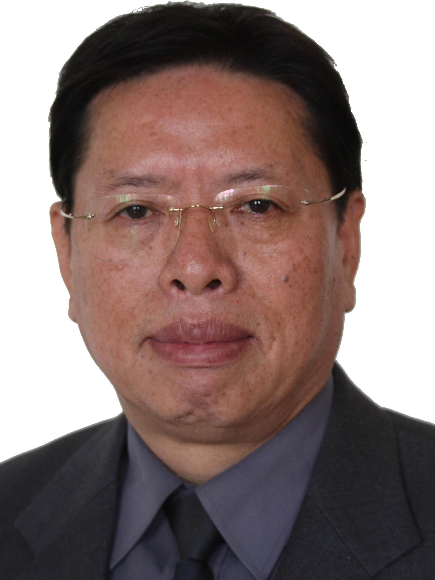
Chen Wufan
Back
Chen Wufan: Chief Scientist of “973 Project”
Basic Information

Name: Chen Wufan
Type of Supervisor: Ph.D. supervisor
Research area: Medical Imaging and Image Analysis
Email: chenwf@smu.edu.cn
Academic Tenure:Standing director of the Chinese Society of Biomedical Engineering, Vice-chairman of China Society of Image and Graphics, Vice-director of Teaching Guiding Committee of the Ministry of Education for Biomedical Engineering, Member of State Key Laboratory of Pattern Recognition, Vice-chairman of Guangdong Society of Biomedical Engineering, and IEEE Senior member.
Research summary
The appearance of large scale medical imaging equipment in 1970s has brought a revolutionary change to clinical and basic medicine, and meanwhile, led to the founding of a new science, “Medical Imaging and Image Analysis.” In 1980s the science of medical imaging and image analysis developed rapidly, and corresponding world-renowned academic journals sprang up. Professor Chen Wufan perceived the great potential of this new science and became the first to engage in medical imaging and image analysis. In 1987 in former First Military University he founded a research center for cross-disciplinary study of information science and life science, which remains a top research institution in China. Prof. Chen has devoted to talent training and cross-disciplinary research that integrate study of natural science, engineering and medicine for over 20 years. Centering on the field of “Medical Imaging and Image Analysis” with significant achievements, Prof. Chen worked out a method of parameter automatic correction based on priori information, and a strategy of two-phase search optimization, which are important innovations of the Optimization theory. He was the first to notice the link between the randomness of information generation and the fuzziness of visual assessment: he advanced the idea of “fuzzy random modelling” for medical image reconstruction and analysis, and the idea of “generalized fuzzy set” (GFS) with an awareness of its transformation relationship to “fuzzy set”; besides, he worked out different models to deal with different imaging and analysis problems. Prof. Chen not only undertakes basic theoretical research, but is also concerned with practical problems. He has deep insights into the practical use of medical imaging science, for instance, MRI, PET, CT, etc. He has applied for patents for some of his imaging techniques, and is also working some companies in China on relevant area. One of his findings, the PACS software, has entered the stage of practical implementation: this software has been used by 20 hospitals in China to ensure their daily working procedure. It is important to emphasize that Prof. Chen applied his findings to form fPAX in the PACS system, which enables the PACS system to perform digital and quantitative clinical diagnosis, and at the same time has strong image analysis capability.
In conclusion, in the past decades Prof. Chen has founded the new science of “Medical Imaging and Image Analysis” as a cross-interdisciplinary study of natural science, engineering and medicine—a difficult journey of forming something out of nothing. Now, he continues to play an important part: as director of Committee of China Medical Imaging and IEEE Senior Member, he works to foster academic exchange between scholars at home and abroad, which helps secure China’s important place in the world in terms of medical imaging and image analysis; as director of Guangdong Key Laboratory of Medical Image, he carries on innovative research on medical imaging and image analysis; as director of Research Center of Digital Medical Equipment, Ministry of Education, he is devoted to pushing technological inventions towards industrial transformation; as vice-director of the Teaching Guiding Committee of the Ministry of Education for Biomedical Engineering, he works heart and soul for more standard undergraduate education, and one of his courses, “Modern Medical Imaging Techniques,” is granted the title “State-level Course of Fine Quality.” Up till now, he has turned out 23 Ph.D. students, 57 Master’s students and 5 post-doctoral students, all of whom are playing a pivotal role in their working units.
Awards and Titles:
1. Guangdong Science and Technology Award, First Prize: “The Study and Application of Recognition System of Tumor Types Based on Image Content” (NO. B07-0-1-02-R01), 2012: the lead author.
2. Guangdong Science and Technology Award, Second Prize: “The Quantitative Measurement System of Heart Function Parameter Based on Series Image” (NO. B07-0-2-05-R01), 2009: the lead author.
3. China National Award for Technological Invention, Second Prize: “The Study of New Medical Imaging and Image Analysis Based on Fuzzy Random Modelling” (NO. 2007-F-235-2-03-201), 2007: the lead author.
4. China National Award for Technological Invention, First Prize; National Science and Technology Nomination Award, Ministry of Education: “The Study and Application of fPAX” (2005-119), 2005: the lead author.
5. Science and Technology Award for Chinese university, Second Prize: “The Theory of General Fuzzy Set and Its Application in Image Processing” (2000-149), 2000: the lead author.
Monograph: Wavelet Analysis and Its Application in Image Processing (lead author). Beijing: Science Press, 2002.
Major papers:
|
Title
|
Journal
|
Impact factor
|
|
Prostate Segmentation based on Variant Scale Patch and Local Independent Projection
|
IEEE Transactions on Medical Imaging
|
3.77
|
|
Hierarchical Patch-Based Sparse Representation—A New Approach for Resolution Enhancement of 4D-CT Lung Data
|
IEEE Transactions on Medical Imaging
|
3.77
|
|
Segmentation of brain magnetic resonance angiography images based on MAP–MRF with multi-pattern neighborhood system and approximation of regularization coefficient
|
Medical Image Analysis
|
4.292
|
Ongoing Research Projects:
|
Name of Project
|
Level of Project
|
Support Fund (10000 RMB)
|
|
Research on major scientific instruments and key techniques for small animal multi-model molecular imaging
|
State-Level
|
300
|
|
The Study of New Techniques of segmenting prostate tumor image based on joint characteristics of multi-model image.
|
State-Level
|
85
|
